

October 9, 2026
What a Scan Report Says Matters: Notes From the South West Imaging Training Academy Endometriosis and Adenomyosis Day
Dr Jodie Hughes, Founder, Endometriosis South Coast · October 2026
I was invited to join the NHS South West Imaging Training Academy for its endometriosis and adenomyosis training day, and to speak about the lived experience of endometriosis. It was a room full of sonographers, clinicians and trainers who give up their time to get better at finding conditions that are too often missed. That alone says a lot about where ultrasound care is heading.
This piece brings together what I spoke about and what I learned from the other speakers, because so much of it matters to our community.
Why the words on a report matter
My talk focused on language: how we speak to people with endometriosis and adenomyosis, and the damage a “normal” scan report can do.
Endometriosis is a whole-body, inflammatory condition that affects the reproductive system, with effects that reach well beyond the pelvis [1]. Many people wait years for a diagnosis. The most recent UK-wide figure from Endometriosis UK is an average of 9 years and 4 months [2]. Ultrasound is often one of the first tests people have on that journey, and for many it is the first time they hope someone will finally see what they have been describing.
When that report comes back saying “normal,” the clinical meaning is usually narrow: nothing was seen on that scan, on that day, with that equipment and that level of expertise. But that is rarely how it lands. For a patient who has been told for years that their pain is just a bad period, “normal” can sound like confirmation that there is nothing wrong, or worse, that they have been making it up. It can delay referral, end conversations with a GP, and teach people to stop asking for help.
National guidance is clear that this should not happen. NICE advises that endometriosis should not be ruled out if an examination and ultrasound scan are normal, and that referral may still be needed even with a normal scan [3]. A scan can show us a great deal, but it cannot show us everything, and the report needs to say so.
A normal scan is not the same as a normal experience.
New national guidance and a tiered approach
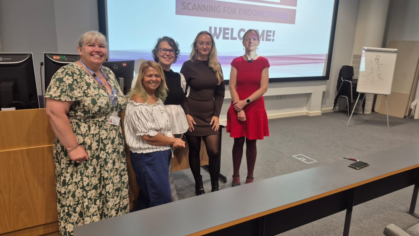
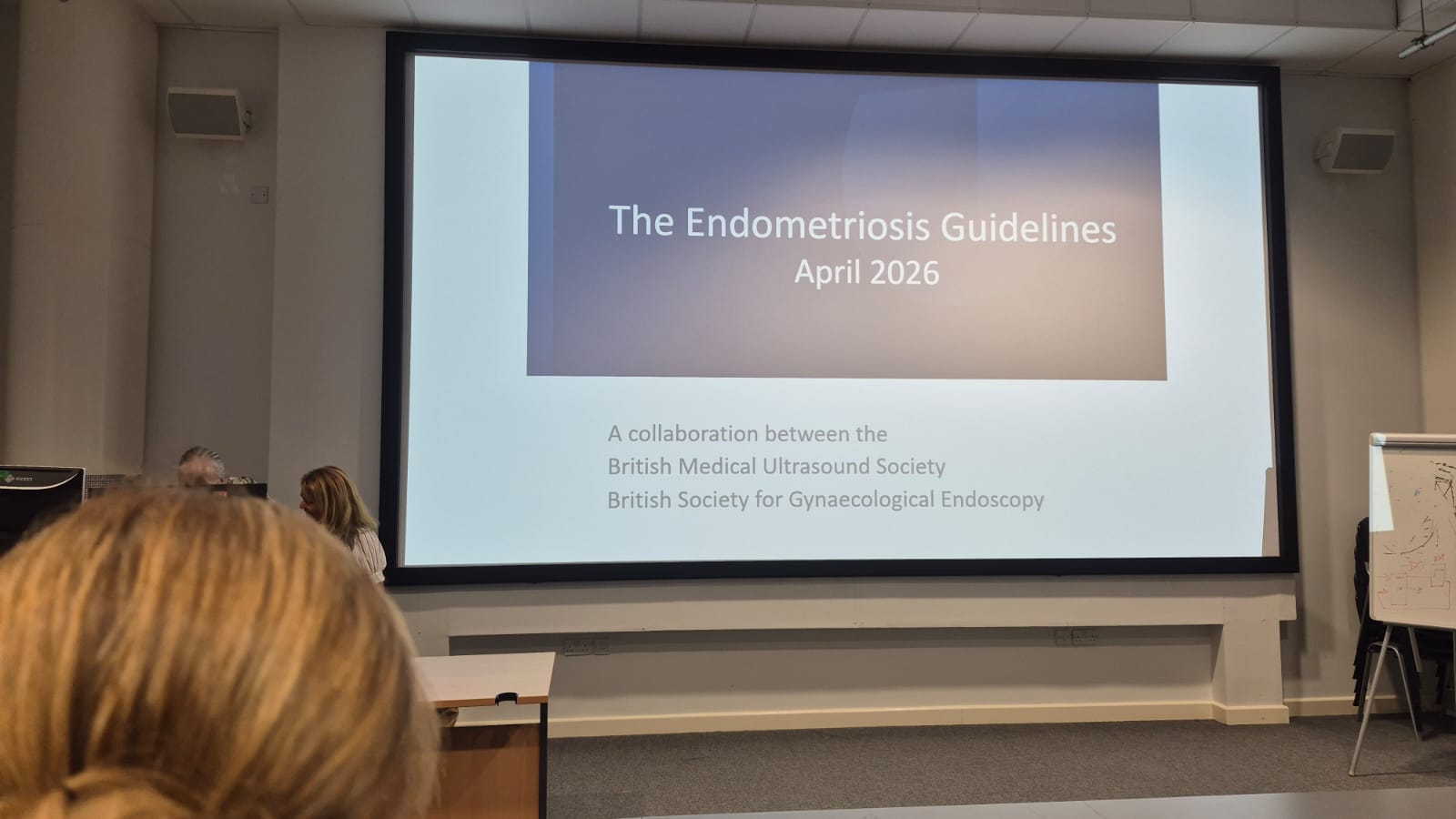
A theme throughout the day was the new guidance on ultrasound assessment of endometriosis from the British Medical Ultrasound Society (BMUS), developed with the British Society for Gynaecological Endoscopy (BSGE). Launched in April 2026, it sets out a tiered framework of expertise and a structured anatomical approach to assessment, with the stated aim of improving detection and helping to reduce diagnostic delay [4, 5].
Speakers stressed that everyone performing pelvic ultrasound should be able to carry out a level 1 scan, with clear routes to more specialist assessment when needed. Borsha Sarkar, speaking on the BSGE and BMUS guidelines, also made the point that the more complicated findings need explaining, both to referrers and to patients, rather than being left as technical shorthand.
Building an invested workforce
Borsha also spoke about building an invested workforce, using the example of a women’s health hub that wanted to set up an ultrasound clinic inside the hub. They trained a nurse and a GP, and worked with a Care Quality Commission (CQC) registered provider using part-time sonographers and students. The speaker reported a return of £8 for every £1 spent.
It raised a question we will be taking forward locally: can our Integrated Care Board (ICB) make a plan for neighbourhood care in Hampshire and the Isle of Wight that includes community diagnostics? Bringing skilled ultrasound closer to where people live could make a real difference to how quickly people with suspected endometriosis or adenomyosis are seen.
A simple approach to scanning for endometriosis
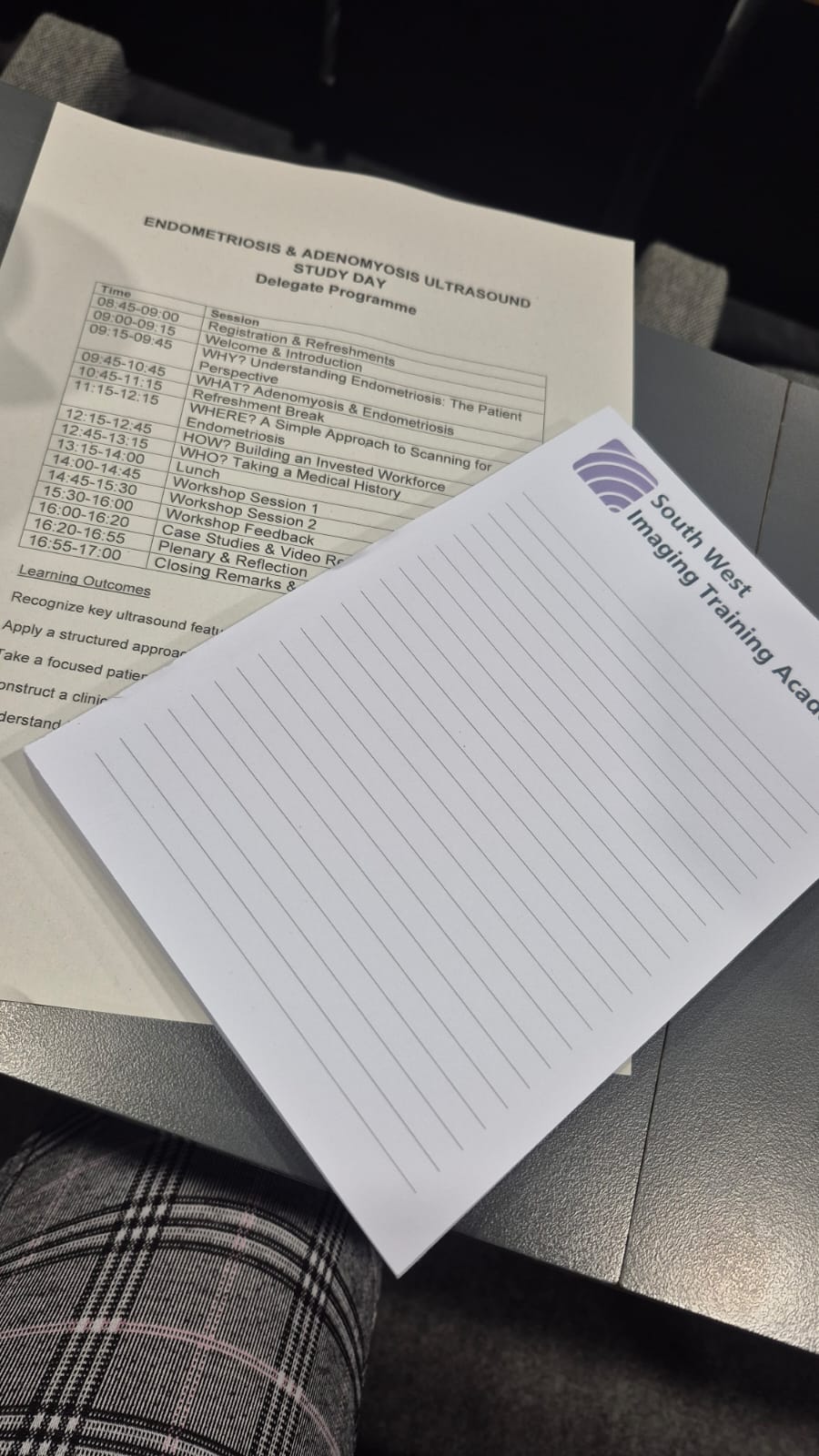
Hania Rytkewska-Siggers gave a practical session titled “A simple approach to scanning endometriosis.” The biggest focus was the sliding sign, a real-time technique where gentle pressure with the probe shows whether the uterus and ovaries slide freely over the bowel and surrounding structures. International consensus guidance describes this as part of a systematic approach to scanning when endometriosis is suspected [6].
She spoke at length about reduced mobility of the ovaries, bowel, uterus and pouch of Douglas (the space between the uterus and the rectum). When organs that should move freely are stuck, that points towards adhesions and disease. She also returned to “site-specific tenderness,” meaning pain that is reproduced when a particular area is pressed during the scan, and to the importance of using clear language when reporting it.
Letting the patient’s history guide the scan
Mrs Alice Bevan, from the BSGE-accredited endometriosis centre in Somerset, asked a question every scan should start with: what does the patient’s history tell you about how to perform this scan, and where can it point you? Symptoms such as pain with bowel movements, painful sex or pain passing urine can tell a sonographer where to look more carefully. For patients, this is a strong reason to share your full symptom history, and a reminder that being asked about it is part of good care.
Describing what is seen, not just labelling it
Dr Susanne Johnson, who also runs an educational YouTube channel, spoke about thickening of the uterosacral ligaments, the ligaments that support the uterus. Her point was that a thickened ligament should not simply be reported as “abnormal” with no further explanation. Thickening can have several causes, including endometriosis, inflammation, fibrosis, adhesions and pelvic inflammatory disease (PID), while a nodule on the ligament points to endometriosis. Describing what is actually seen helps the next clinician make sense of it, and helps the patient understand what happens next.
Adenomyosis is often missed
Adenomyosis came up repeatedly. One speaker shared that around 1 in 5 hysterectomy specimens show adenomyosis, and that many of these had been reported on scan as diffuse fibroid change rather than adenomyosis. The message was that diffuse change of this kind should prompt careful consideration of adenomyosis. For our community, that means it is reasonable to ask whether adenomyosis has been considered if a report describes diffuse or fibroid-like changes alongside symptoms such as heavy, painful periods.
How new practice can be introduced
The day closed with practical advice on bringing these skills into services:
- Create a small trained team rather than trying to change everything at once.
- Build in supervised scanning, case reviews and ongoing learning.
- Be clear about who works at each level for specialist scanning, so patients are routed to the right expertise.
What this means for our community
I came away hopeful. The people in that room want to get this right, and the new guidance gives them a shared framework to do it. But the most important change may be the simplest: making sure every report is written with the person reading it in mind. If you have been given a “normal” scan but your symptoms continue, that does not mean your pain is not real, and it does not mean the search for answers should stop. You are entitled to ask what the scan could and could not show, and whether further assessment or referral is needed.
Thank you to the South West Imaging Training Academy for inviting Endometriosis South Coast, and to every speaker and delegate for the work they are doing.
References
- Taylor HS, Kotlyar AM, Flores VA. Endometriosis is a chronic systemic disease: clinical challenges and novel innovations. The Lancet (2021), 397(10276): 839-852. doi: 10.1016/S0140-6736(21)00389-5
- Cysters & Endometriosis UK. Disbelieved, Dismissed, Delayed: Endometriosis diagnosis inequalities in ethnically diverse communities (March 2026), citing Endometriosis UK, The state of endometriosis care across all 4 nations (2026). endometriosis-uk.org
- National Institute for Health and Care Excellence. Endometriosis: diagnosis and management (NG73), 2017, updated November 2024. nice.org.uk/guidance/ng73
- British Medical Ultrasound Society and British Society for Gynaecological Endoscopy. Guidance for Ultrasound Assessment of Endometriosis (2026). bmus.org
- British Medical Ultrasound Society. Guidance for Ultrasound Assessment of Endometriosis (news release, 17 April 2026). bmus.org/mediacentre/news/guidance-for-ultrasound-assessment-of-endometriosis
- Guerriero S, Condous G, van den Bosch T, et al. Systematic approach to sonographic evaluation of the pelvis in women with suspected endometriosis, including terms, definitions and measurements: a consensus opinion from the International Deep Endometriosis Analysis (IDEA) group. Ultrasound in Obstetrics & Gynecology (2016), 48(3): 318-332. doi: 10.1002/uog.15955